Ectoderm – Surface Ectoderm – Neural tube – Neural crest
In my previous post, I wrote about neurulation (i.e. the development of the neural tube and neural crest) and in the list at the end I wrote a bit about what those structures can develop into. There's not a lot more to develop on that, which means less reading for you and less typing for me :D
The anterior pituitary is one of the structures that develops from the surface ectoderm, as previously listed. It does this by folding inwards to create a structure known as Rathke's pouch. Rathke's pouch then detaches and moves next to the infundibulum of the developing brain (it's essentially a protrusion on the base of the brain, to my understanding) to form the anterior part of the pituitary gland.
I'm just going to change topics a bit here to talk about the oral and cloacal membranes. One thing I forgot to mention in my last post is that the mesoderm doesn't completely separate the endoderm and ectoderm. Instead, there are two places where the endoderm and ectoderm remain directly in contact. One place is near the cranial end, and is known as the oral membrane. The other place is near the caudal end, and is known as the cloacal membrane. The oral membrane eventually ruptures through and forms the oral cavity, whereas the cloacal membrane forms the anal, genital and urinary openings when it ruptures through.
The reason I bring this up now is because the development of Rathke's pouch and thus the anterior pituitary also highlights the fact that the oral membrane is behind the mouth. Confused? Well, since the oral membrane is the location where the ectoderm and endoderm meet, and it ruptures through to form the oral cavity, anything past the oral membrane is formed from endoderm (like the lining of your oesophagus and digestive system). However, the anterior pituitary, which is formed from cells lining the mouth, is ectodermal in origin. Hence the mouth, and the lining of the mouth, must be in front of the oral membrane.
Now I've spoken about surface ectoderm, I'm going to move on to neural tube ectoderm. As mentioned in my previous post, it's responsible for the formation of the central nervous system- that is, the brain and spinal cord. The neural tube is expanded at the cranial end, which forms the brain. The canal that runs through the neural tube eventually becomes the ventricles of the brain (cerebrospinal fluid flows through the ventricles). Motor neurons are located on the ventral side of the canal whereas sensory neurons are located on the dorsal side.
The canal is made up of grey matter and white matter, named due to their appearance. Grey matter is comprised of nerve cell bodies, while white matter is comprised of myelinated axon sheaths (myelin looks white, hence the name). In the part of the canal that eventually becomes the spinal cord, grey matter is found more on the inside lining the canal, while white matter is found on the outside. This situation is reversed in the brain.
Finally, a quick word on the neural crest ectoderm. There's not much I can expand on here, other than giving some more examples of things that the neural crest ectoderm eventually becomes. Aside from the sensory epithelia of the eyes, ears and nose, peripheral nervous system and adrenal medulla (yup, I copy-pasted that list from the previous post and edited in the word "and"), the neural crest cells also develop into Schwann cells (oligodendrocytes which form the myelin sheath in the peripheral nervous system), the pia and arachnoid meninges (which are collectively known as "leptomeninges"), the dentine of the teeth, the cornea of the eyes and melanocytes (the cells that produce melanin). The neural crest also forms much of the cartilage, muscle and so forth in the head that were previously thought to have developed from mesoderm. And speaking of the head...
Development of the Head
Many structures in the head are simply continuations of structures in the rest of the body. For example, as previously mentioned, the neural tube continues up into the head to become the brain. The vertebral column also continues up to form the base of the skull. Many structures, such as the cranial vault, pharyngeal arches (precursors for structures such as the jaw, larynx and other structures in the neck) and sensory placodes (i.e. the eyes, ears and nose) develop via interaction between the neural tube or neural crest with the surface ectoderm.
Mesoderm – Paraxial – Intermediate – Lateral plate
I don't think I have much to say on the mesoderm. That being said, I didn't think that I had much to say on the ectoderm, either, and I wrote six paragraphs on it, not counting my whiny introductory paragraph.
I'll start with the lateral plate mesoderm, because I'm fairly sure my first post pretty much covered this. I did promise that I'd talk about the somatopleure and visceropleure a bit more, though, which was kind of stupid of me seeing that I don't really have much more to say about them. Both are responsible for the formation of smooth muscle, connective tissue and vasculature, but the somatopleure is responsible for the stuff associated with the epidermis of the body wall and limbs and the visceropleure is responsible for the stuff associated with the epithelia of the gut.
Now I'll talk a bit about the paraxial mesoderm, and this time I'm fairly sure that I do have something to say about it that I haven't said before: namely, the idea of the epimere and hypomere. Some cells of the dermatome and myotome remain put and are known as the epimere, while others migrate into the somatopleure and are known as the hypomere. (I'm not sure about sclerotome, though- maybe this is why last year they told us that sclerotome only forms the axial skeleton?) Cells of the epimere become the dermis and muscles of the back, while cells of the hypomere become the dermis and muscles of the body wall and limbs.
Before I get onto how this relates to nerve supply to different organs (because yes, this epimere/hypomere stuff relates to nerve supply to different organs), I'm first going to have a quick talk about typical spinal nerves. Behold, this picture that I so rudely took from http://thegoofyanatomist.weebly.com/uploads/3/0/9/9/30995885/1422050582.png:
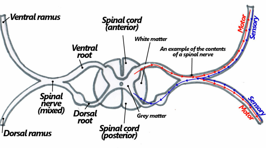
As I've mentioned before, sensory nerves are found on the dorsal (posterior) side of the cord, whereas motor nerves are found on the ventral (anterior) side of the cord. (Just in case you want to know, that bulge on the dorsal side is known as the "dorsal root ganglion," and it contains a cluster of sensory nerve cell bodies.) Sensory and motor neurons eventually meet to form a mixed spinal nerve, as you can see on the right side of the diagram. Mixed spinal nerves branch out into two divisions: the ventral rami and the dorsal rami. I'll get to them in a bit. Some nerves, specifically the nerves between T1 and L1, also have another "white ramus" which leads to a sympathetic ganglion of the sympathetic trunk. From here sympathetic nerves run to the body wall via the grey ramus, or to the viscera via visceral branches.
Now I'm going to get back to those ventral and dorsal rami. While the cells of the hypomere migrate, they take their own nerve supply with them. This nerve supply is the ventral rami. Hence, the epimere is supplied by the dorsal rami, while the hypomere is supplied by the ventral rami.
For some reason, when we were learning about this during the lecture, I had a mental image of muscle cells (representing myotome) marching to the ventral side of the embryo dragging nerves behind them. No, seriously, here's the picture that I doodled during the lecture:

To top it off, I imagined them migrating to the tune of the Adventure Line music from The Stanley Parable (which, by the way, is also my ringtone. So now you know who to glare at if it goes off in lectures).
There! Now you're never going to be able to forget this, even when you desperately want to! Muahahaha!
Limb Development
We didn't really cover limb development in that much detail (in fact I'm fairly sure the lecturer pretty much skipped over the slide relating to limb development as we were going overtime), so all I can really tell you are the basics. Limbs develop from the somatopleure, which as I've mentioned several times, is the layer of the lateral plate mesoderm that associates with the body wall. The muscle and skin in this area is from the hypomere and is therefore supplied by ventral rami. Limb development starts from little limb buds that begin to form in the 4th week, which induce the proliferation of underlying mesoderm to create the limb. Formation of limbs are proximodistal (i.e. from the "close" end of the limb to the "far" end).
No comments:
Post a Comment